Adult acquired flatfoot
and tibialis posterior tendonopathy
Imaging
The diagnosis of adult acquired flatfoot is made clinically. If there is no controversy about diagnosis or management we do not routinely obtain radiographs. However, in many patients there is a need to assess the joints and alignment for treatment planning, and in some there may be the possibility of inflammatory arthropathy or a tarsal coalition.
The standard plain films are:
- standing hindfoot alignment view – we use a technique slightly modified from that of Saltzman and el-Khoury (1995) to improve the visualisation of the ankle. Many people refer to this as the Cobey view. It shows the hindfoot alignment and any tilting in the ankle mortise
- standing lateral foot view – shows peritalar and talometatarsal alignment and allows assessment of arthritis
- standing dorsoplantar foot view – as above
On these views we assess the following parameters:
- tibiotalar and tibiocalcaneal alignment
- peritalar malalignment using:
- lateral talometatarsal (Meary’s) angle
- dorsal talometatarsal angle
- talonavicular coverage angle
- cuneometatarsal alignment in lateral and dorsoplantar projections
- other deformities such as hallux valgus
- presence of arthritis, accessory navicular or tarsal coalition
- bone stock
- any previous surgery
|
|
|
Standing hindfoot alignment view
Calcaneal valgus angle should be 5deg
|
Standing lateral view.
Meary's angle (marked) should be <10deg |
Standing dorsoplantar view.
Dorsoplantar talo-first metatarsal angle (left) should be <10deg
Talonavicular coverage angle (right) |
If we are planning surgery, or if there is any other need to assess the tendons and ligaments, we obtain an MR scan. Ultrasonography has also been described and is probably of equal accuracy to MRI, provided expertise and suitable equipment are available.
CT shows bony changes well, but does not show soft tissues clearly.
|
| Transverse MRI showing split in tibialis posterior tendon |
|
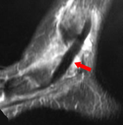
| Sagittal MRI showing thickened tibailis posterior tendon |
MR shows:
- tibialis posterior tendon and pathology
- other long flexor tendon pathology – FHL tendonopathy is sometimes seen in association with tibialis posterior tendonopathy
- the deltoid and, to some extent, the spring ligament
- intra-articular lesions in the ankle and other joints
- bone oedema, sometimes seen in the lateral calcaneum in lateral impingment, or in the navicular
Conti et al (1992) classified the MR appearances:
- type 1: longitudinal splits without tendonopathy
- type 2: swelling and degeneration
- type 3: replacement of tendon substance with scar
Conti et al found that the MR appearances were a better guide to outcome than surgical findings. In particular tendon transfers were significantly more successful in type 1 tendons, but tendons graded type 1 by the surgeon were graded type 2 by MR in 10/17 patients - intra-operative assessment may lead to inappropriate choice of treatment.