Ankle fractures
Anatomy and biomechanics
The ankle is a modified hinge joint between the tibial plafond, medial and lateral malleoli proximally and the talus distally.
The inferior tibiofibular and subtalar joints are also intimately related to ankle function.
The ankle joint capsule is reinforced by the anterior talofibular (ATFL), calcaneofibular (CFL) and posterior talofibular ligaments (PTFL) laterally, and by the deltoid ligament medially, of which the deep tibiotalar part is the most important for ankle stability.
There are also anterior (ATFL), interosseous and posterior ligaments of the inferior tibiofibular joint and a posterior transverse band, the posterior intermalleolar ligament.
The subtalar joint is stabilised by the lateral, interosseous and cervical talocalcaneal ligaments, and by the calcaneofibular and superficial deltoid ligaments and the inferior extensor retinaculum, which cross both ankle and subtalar joints.
 |
 |
 |
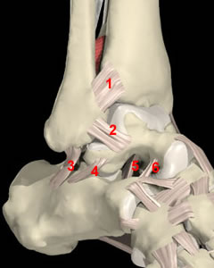
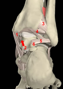
Anterolateral ankle structures
1 Anterior tibiofibular ligament
2 Anterior talofibular ligament
3 Calcaneofibular ligament
4 Lateral talocalcaneal ligament
5 Interosseous talocalcaneal ligament
6 Cervical talocalcaneal ligament
|
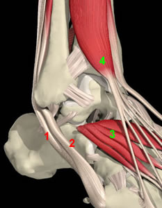
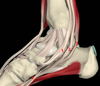
More superficial structures
1 Peroneus longus
2 Peroneus brevis
3 Extensor digitorum brevis
4 Extensor digitorum longus
Notice how the calcaneofibular ligament lies in the floor of the peroneal tendon sheath. Tears of this ligament allow communication between the ankle and the sheath through which contrast or an arthroscope can pass |
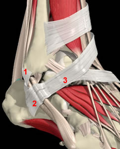
More superficial again
1 Superior peroneal retinaculum
2 Inferior peroneal retinaculum
3 Inferior extensor retinaculum
The superior peroneal retinaculum may be torn in lateral ligament injuries
The inferior extensor retinaculum contributes to subtalar joint stability
|
 |
 |
Medial ankle structures
1 Parts of superficial deltoid ligament
2 Spring ligament
3 Short plantar ligament
The superficial tibiotalar ligament (1 posterior) is intermediate between superficial and deep deltoid |
More superficial structures
1 Tibialis posterior
2 Flexor dgitorum longus
3 Flexor hallucis longus
4 Knot of Henry |
 |
 |
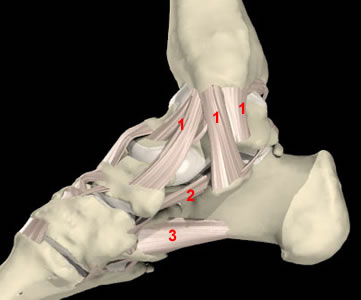
Posterior ankle structures
1 Superficial deltoid ligament
2 Deep deltoid ligament
3 Posterior tibiofibular ligament
4 Posterior intermalleolar ligament
5 Posterior talofibular ligament
6 Posterior talar process/os trigonum
The deep deltoid ligament is the most important structure in the fractured ankle as its integrity determines fracture stability
Tears of the deep deltoid ligament produce the "posteromedial impingement lesion"
The posterior ligaments are well seen at arthroscopy
|
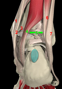
More superficial structures
1 Tibialis posterior
2 Flexor digitorum longus
3 Neurovascular bundle space
4 Flexor hallucis longus
5 Safe space for posterior ankle portals
6 Peroneus brevis
7 Peroneus longus
FHL is the main posterior landmark and can be seen from inside the ankle by manipulating the great toe |
Images are courtesy of Interactive Foot and Ankle (Primal Pictures)
Back to introductory page
Forward to next page (movement)
Forward to next section (pathomechanics and classification)