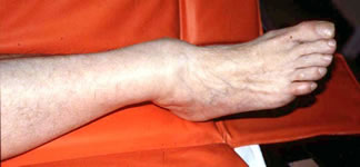
The obvious deformity risks medial skin necrosis. This ankle needs immediate provisional reduction
Patients with ankle fractures normally give a history of a fall or other injury mechanism. Absence of injury should make one consider a neuropathic fracture. Some patients hear a crack which is usually in favour of a major ligament injury or fracture. Other indications of the severity of injury include whether the patient was able to stand on the ankle, to walk, run, continue sport or other activity after injury.
| A general survey of the patient should be undertaken, as meticulous as indicated by the history and severity of injury. There may be obvious malalignment of the ankle. If this is imperilling the skin over the medial side, the ankle needs to be reduced urgently. Inspect the skin and subcutaneous tissues for open fractures, fracture blisters and swelling. | |
The obvious deformity risks medial skin necrosis. This ankle needs immediate provisional reduction |
Palpation of the acutely injured ankle should begin at the proximal fibula. The length of the fibula is palpated for tenderness, swelling or disruption to the bony contour, along with the tibiofibular interosseous area, especially the inferior tibiofibular joint. Clinical evidence of a malleolar fracture should be sought by palpation. If in doubt, stressing the malleoli by pushing the talus medially or externally rotating it may produce pain or displacement, but this is unnecessarily painful if there is already strong evidence of fracture. In undisplaced fractures, carefully examine the soft tissue of the medial side. Tenderness, swelling and bruising here are suggestive of a tear of the deltoid ligament and hence of potential instability. The calcaneum, midfoot and 5th metatarsal should also be examined.
Check the sensation, circulation and pulses. Examine the Achilles, long flexor and peroneal tendons.